
We prepared a dataset consisting of 11,089 haematoxylin-eosin-stained paraffin tissue section images taken from 11 colon adenocarcinoma patients using Leica DMI3000B microscope and Leica Application Suite camera software.
The ethical permission for the study was approved by the ethical committee of the University of Federico II of Naples (protocol number 394/19).
Each series of images cover the whole tissue section.
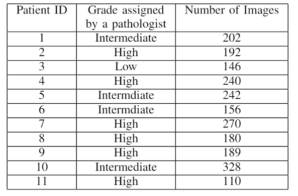
An experienced pathologist, based on the patients’ disease history, did the diagnosis of the degree of colon tumorigenesis.
The grades assigned by the pathologist and the number of images related to each patient are summarized in the following table.
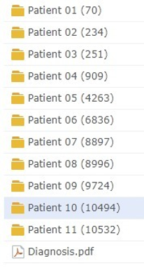
Collected images are divided into directories, each of which refers to a patient (the number in the brackets is for internal reference only). The root directory is reported in the figure on the left.
The selected patients represent advanced pT3 and pT4 stages of adenocarcinoma with neoplastic infiltration to neighbouring tissues, excluding the samples from patients 1 and 3.
The sample from patient 1 was isolated from a liver metastasis, whereas patient 3 had a pathological stage pT1 adenocarcinoma with no metastasis.
The file named “diagnosis.pdf”, placed in the main directory, contains detailed information about patients’ diagnosis.
The dataset can be found here :

To access and download the dataset a password in needed.
If you are reviewing the manuscript that introduces the dataset, you can use the first word in the title of the paper (upper case and lower case as appear). Otherwise, please contact Marco Leo for access information:

Image size is 1824×1368 pixels, in tiff format, and each image requires about 7 MB disk space.